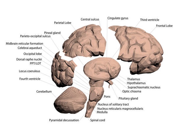
GENERAL APPEARANCE
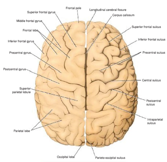
- The cerebral hemispheres are the largest part of the brain; they are separated by a deep midline sagittal fissure, the longitudinal cerebral fissure.
- The fissure contains the sickle-shaped fold of dura mater, the falx cerebri, and the anterior cerebral arteries.
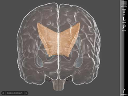
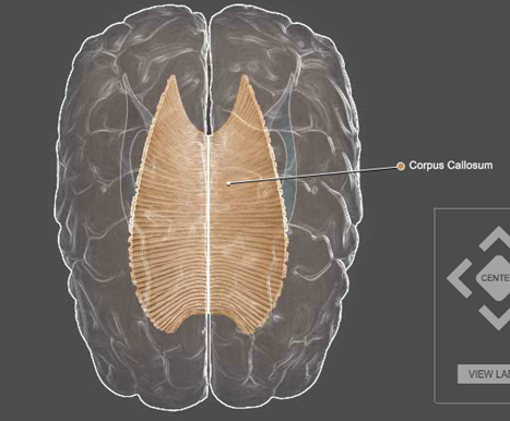
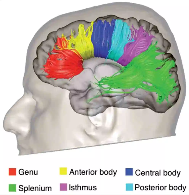
- In the depths of the fissure, the great commissure, the corpus callosum, connects the hemispheres across the midline.
- A second horizontal fold of dura mater separates the cerebral hemispheres from the cerebellum and is called the tentorium cerebelli
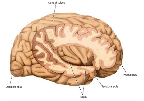
POLES
- Frontal pole
- Occipital pole
- Temporal pole
BORDERS
- Superomedial
- Inferolateral
- Superciliary
- Medial orbital
- Medial occipital
- Inferomedial
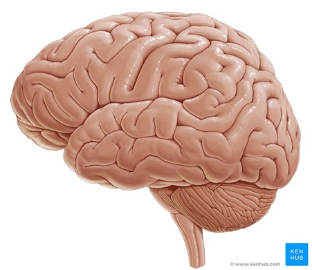
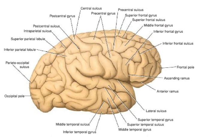
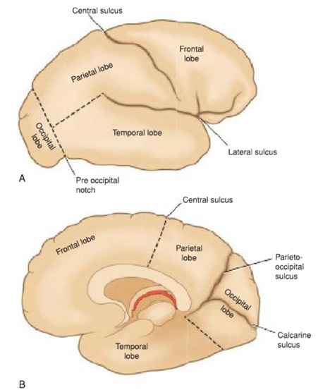
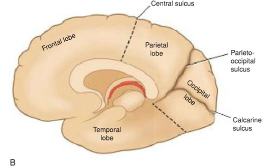
SURFACES
- Superolateral surface
- Medial surface
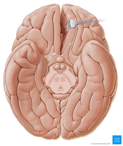
- Inferior surface
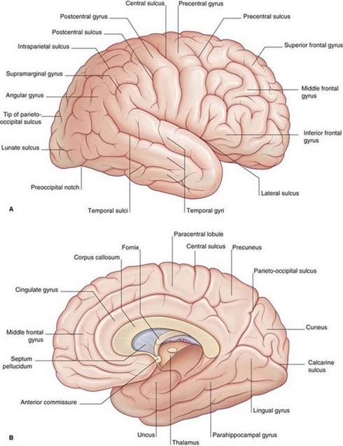
MAIN SULCI
CENTRAL SULCUS:
LATERAL SULCUS
PARIETOOCCIPITAL
CALCARINE
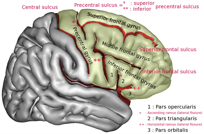
FRONTAL LOBE {3 sulci and 4 gyri}
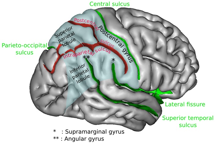
PARIETAL LOBE {2 sulci and 3 gyri}
TEMPORAL LOBE {2 sulci and 3 gyri}
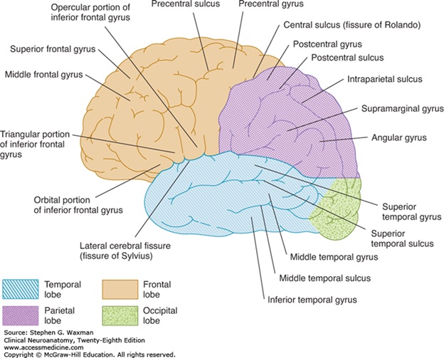
OCCIPITAL LOBE
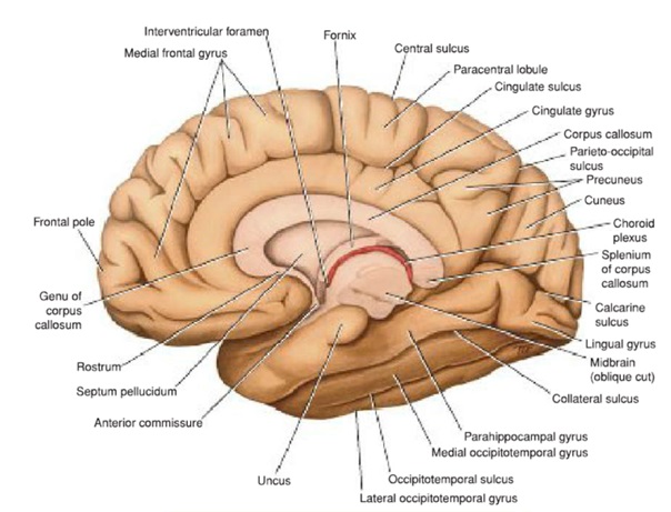
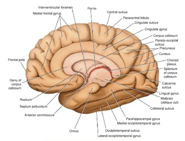
MEDIAL SURFACE
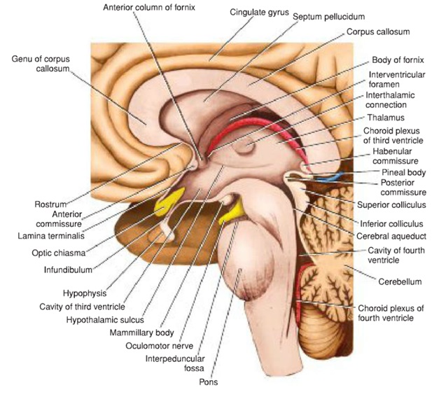
- Corpus callosum
- Cingulate gyrus
- Paracentral lobule
- Precuneus
- Cuneus
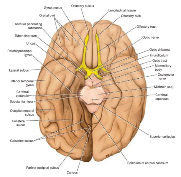
INFERIOR SURFACE
- Collateral sulcus
- Para hippocampal gyrus
- Uncus
- Medial occipitotemporal gyrus
- Occipitotemporal gyrus
- Olfactory sulcus
- Gyrus rectus
- Orbital gyrus
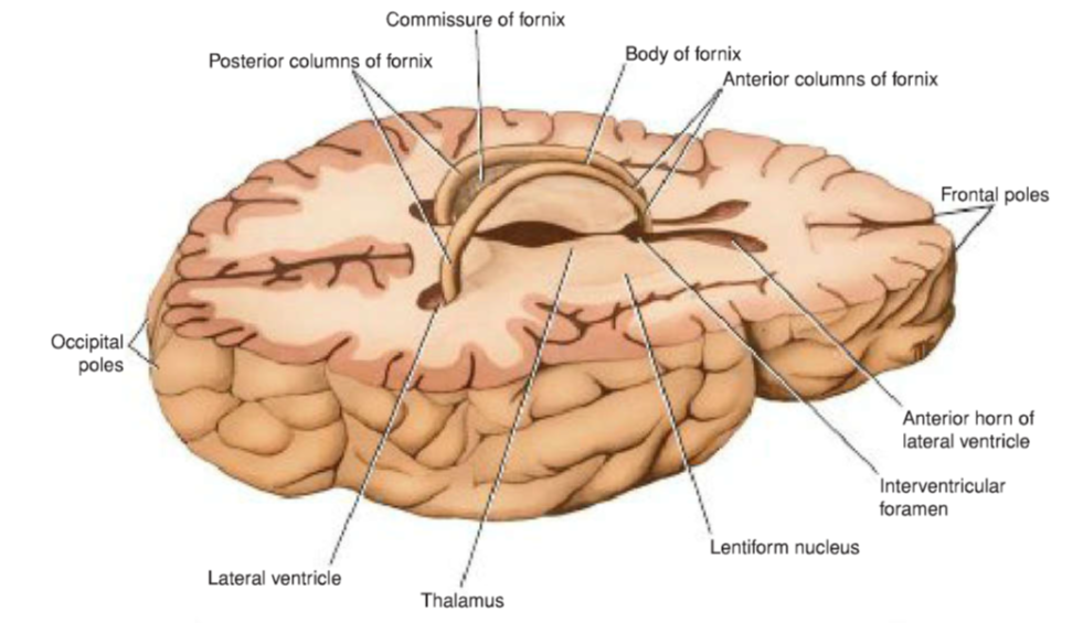
INTERNAL STRUCTURE
- Lateral Ventricles
- Basal Nuclei
- White Matter
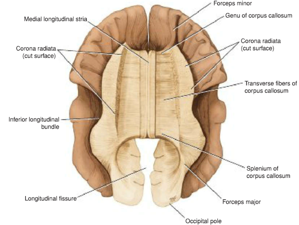
WHITE MATTER
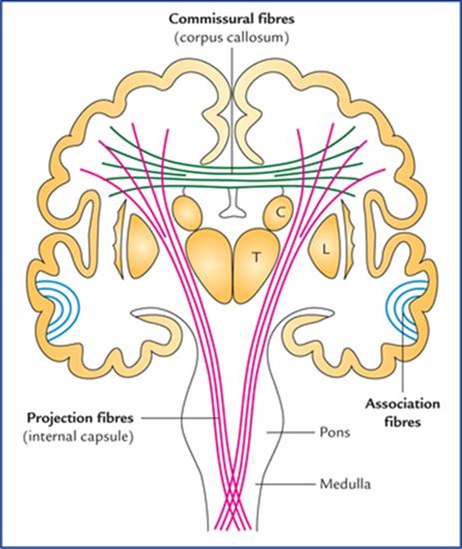
The white matter is composed of myelinated nerve fibers of different diameters supported by neuroglia.
The nerve fibers may be classified into three groups according to their connections:
(1) commissural fibers,
(2) association fibers, and
(3) projection fibers.
COMMISSURE FIBERS
Commissure fibers essentially connect corresponding regions of the two hemispheres
1.corpus callosum
2.anterior commissure
3.posterior commissure
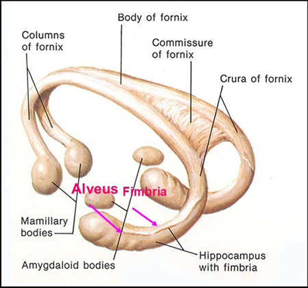
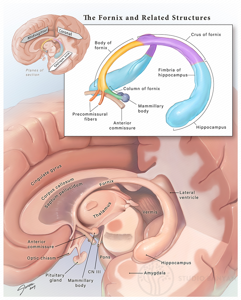
4.fornix
5.habenular commissure
2. ANTERIOR COMMISSURE
3. POSTERIOR COMMISSURE
4. HABENULAR COMMISURE
5. FORNIX
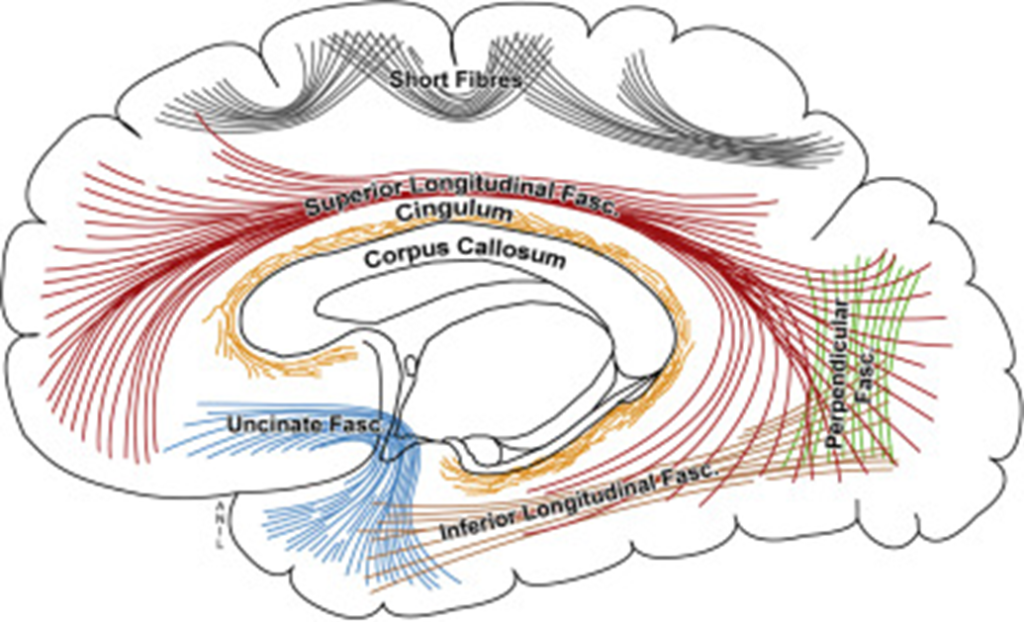
ASSOCIATION FIBERS
Association fibers are nerve fibers that essentially connect various cortical regions within the same hemisphere
1.Short association fibers
2.Long association fibers
1.Uncinate fasciculus
2.Cingulum
3.Superior longitudinal fasciculus
4.Inferior longitudinal fasciculus
5.Fronto-occipital fasciculus
PROJECTION FIBERS
Afferent and efferent nerve fibers passing to and from the brainstem to the entire cerebral cortex must travel between large nuclear masses of gray matter within the cerebral hemisphere
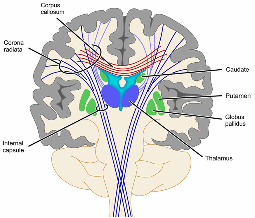
INTERNAL CAPSULE
You can also download other Anatomy Notes from given below buttons
