LEARNING OBJECTIVES
- Identify bones of an articulated hand
Elucidate the salient features of skin of palm and dorsum of hand and discuss its cutaneous innervation
- • Correlate palmar aponeurosis and its septa with palmar spaces
- Enumerate the small muscles of hand with their actions and nerve supply
- Describe the fibrous and synovial flexor sheaths of the hand
- • Explain the carpal tunnel with reference to its formation and contents
- • Analyze the anatomical basis of Dupuytrens contracture, carpal
- tunnel syndrome, trigger finger and tenosynovitis of synovial sheaths of flexor tendons
- • Describe boundaries & contents of spaces of palm
- • Analyze the clinical importance of
Analyze clinical importance of spaces of palm, forearm space of Parona and pulp spaces
• Revisit the insertion of long flexor and extensor tendons
• Describe the blood supply of hand
• Describe the formation of Superficial and Deep Palmar Arches in hand
• Trace the pathway and distribution of radial, median and ulnar nerves in hand
• Identify the muscles, nerves and vessels of hand on prosected specimens and models


Specific Points
1.The scaphoid.
The tubercle is directed laterally , forward and downwards.
Scaphoid Bone Articular Surfaces

Specific Points
2.Lunate:
semiluen shape bone lie medial to scaphoid. Articulate with scaphoid laterally and triquetral medial. Proximally attached with radius.
Lunate Bone Articular Surfaces

3.The Triquetral.
(i) The oval facet for the pisiform
lies on the distal part of the palmar surface,
(ii)The medial and dorsal surfaces are continuous and non-articular.
Triquetrum Bone Articular Surfaces

4.The pisiform.
(i) The oval facet for the triquetral
lies on the proximal part of the dorsal surface,
(ii) The lateral surface is grooved by the ulnar nerve.
Pisiform Bone Articular Surface

The trapezium,:
(i) The palmar surface has a
vertical groove for the tendon of the flexor carpi radialis.
(ii) The groove is limited laterally by
the crest of the trapezium
(iii) The distal surface bears a concavoconvex articular surface for the base of the first metacarpal bone.
Trapezium Bone Surfaces and Articulation

The trapezoid.
(i) The distal articular surface is
bigger than the proximal, (ii) The palmar nonarticular surface is prolonged laterally.

The capitate.
The dorsomedial angle is the
distal-most projection from the body of the capitate. It bears a small facet for the 4th metacarpal bone.

The hamate.
The hook projects from the distal part of the palmar surface, and is directed
laterally.

Carpal Tunnel

Metacarpals

Metacarpals


Phalanges of the Hand

EXTRINSIC MUSCLES OF HAND

Muscles of the Forearm Move the Wrist Hand Thumb and Fingers

Extrinsic Hand Muscles

Intrinsic muscles of hand

Intrinsic Hand Muscles


Intrinsic Hand Muscles

Thenar Muscles

Abductor Pollicis Brevis

Flexor Pollicis Brevis

Opponens Pollicis

Adductor Pollicis

Hypothenar Muscles

Opponens Digiti Minimi

Abductor Digiti Minimi

Flexor Digiti Minimi Brevis

Intermediate Muscle

The first lumbrical arises from the radial side of the tendon OF FBP for the index finger .
The second lumbrical arises from the radial side of the tendon for the
middle finger.
The third lumbrical arises from contiguous sides of the tendons for the middle and ring fingers.
The fourth lumbrical arises from the
contiguous sides of the tendons for the ring and little fingers.
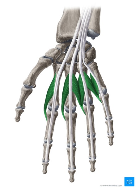
Nerve Supply:
1.The first and second lumbricals by the median nerve
(C8, Tl).
2.The third and fourth lumbricals by the deep branch of the ulnar nerve (C8, Tl).
Actions:
The lumbrical muscles flex the metacarpophalangeal joints, and extend the interphalangeal joints of the digit into which they are inserted.Deep to the lateral two tendons of flexor digitorum
Lumbricals


Palmar Interosei

1.First palmar interosseous muscle from the medial side of the base of the first metacarpal bone.
2.Second palmar interosseous muscle from the medial half of the palmar aspect of the shaft of the second metacarpal bone
3. Third arises from the shaft of the palmar aspect of 4th metatarsal
4. Fourth from the lateral part of palmar aspect of shaft of fifth metatarsal.
Insertion:
Each muscle is inserted into the dorsal digital expansion of one digit. It may also be attached to the base of the proximal phalanx of the same digit. The digits
into which individual palmar interossei are inserted
are as follows.
1.First muscle: Medial side of thumb.
2.Second muscle: Medial side of the index finger.
3.Third muscle: Lateral side of the fourth digit.
4. Fourth muscle: Lateral side of the fifth digit.
Note that the middle finger does not receive the
insertion of any palmar interosseous muscle.

Nerve supply & action
- Nerve Supply:
All palmar interossei are supplied by the deep branch of the ulnar nerve (C8, Tl).
- Actions:
- All palmar interossei adduct the digit to which they are attached towards the middle finger. In addition,they flex the digit at the metacarpophalangeal joint and extend it at the interphalangeal joints.
DORSAL INTEROSEI
- 1.First dorsal interosseous:
Shafts of first and second metacarpals.
- 2.Second dorsal interosseous:
Shafts of second and third metacarpals.
- 3.Third dorsal interosseous:
Shafts of third and fourth metacarpals.
- 4.Fourth dorsal interosseous:
Shafts of fourth and fifth metacarpals.
DORSAL INTEROSEI
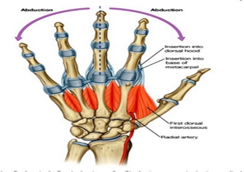
Insertion
- Each muscle is inserted into the dorsal digital expansion of the digit; and into the base of the proximal phalanx of that digit. The digits into which individual muscles are inserted are as follows:
- 1.First: Lateral side of index finger.
- 2.Second: Lateral side of middle finger.
- 3.Third; Medial side of middle finger.
- 4.Fourth: Medial side of fourth digit.
- Note that the middle linger receives interosseous muscle on either side; and that the first and fifth digits do not receive any insertion

NERVE SUPPLY & ACTION
- Nerve Supply:
All dorsal interossei are supplied by the deep branch of the ulnar nerve (C8, Tl).
- Actions:
All dorsal interossei cause abduction of the digits away from the line of the middle finger. This movement occurs in the plane of palm in contrast to the movement of thumb where abduction occurs at right angles to the plane of palm. Note that movement of the middle finger to either medial or lateral side constitutes abduction. Also note that the first and fifth digits do not require dorsal interossei as they have
their own abductors. In addition (like the palmar interossei), the dorsal interossei flex the metacarpophalangeal joint of the digit concerned and
extend the interphalangeal joints.

