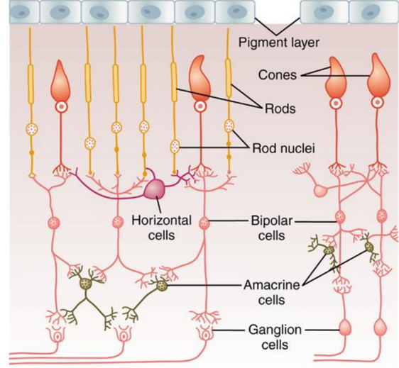
- Rods and cones
- Horizontal cells
- Bipolar cells
- Amacrine cells
- Ganglion cells
- Interplexiform cells
- Rods and cones– Photoreceptors
- Horizontal Cells—Inhibitory cells
- Bipolar Cells– Depolarising/Hyperpolarising
- Amacrine Cells—30 types
- Ganglion cells—W, X and Y cells
Visual Pathway in Retina
- Cone vision– foveal portion of retina
– 3 cell pathway
– Cones → bipolar cells → Ganglion cells
– New and fast system - Rod vision– peripheral retina
– 4 cell pathway
– Cones → bipolar cells → amacrine cells →
Ganglion cells
– New and fast system
Neurotransmitters
- Glutamate
- Amacrine cells secrete 8 types of NT
– GABA
– Glycine
– Dopamine
– Acetylcholine
– Indolamine
Signal Transmission in Retina
- Ganglion cells and amacrine cells
– True action potential - Amacrine cells, horizontal cells and
Photoreceptors
– Electrotonic conduction
– Graded Potential
Photoreceptors
- Rods and cones
- Transmits signals to outer plexiform layer
- Synapse with bipolar and horizontal cells
Horizontal cells
- Transmits signals horizontally to bipolar cells
- In the outer plexiform layer
- Transmits inhibitory signals laterally
- Plays role in lateral connectivity along with
amacrine cells - Synapse with dendrites of bipolar cells and
cell bodies of rods and cones - Obeys phenomena of lateral inhibition
- Lateral inhibition– transmission of visual
patterns with proper visual contrast - Light strikes eye– central area excited
- Outer zone–inhibited by lateral inhibition via
horizontal cells - amacrine cells also contribute in lateral
inhibition - Enhancement of visual contrast

Bipolar Cells
- 2 types
– Hyperpolarizing cells
– Depolarizing cells - 1
st order neuron in visual pathway - Cells respond to glutamate released by rods
and cones in their specific manner
- Bipolar (Depolarizing) cells takes excitation
from rods and cones - Bipolar (Hyperpolarizing) cells takes signals
from horizontal cells - Half of the cells transmit positive signals and
rest half sends negative signals - Contributes in lateral inhibition and provides
contrast border in the image
Amacrine Cells
- 30 types
- Functions
- Conduction Pathway (Rod vision)
- Offset image
- Respond to change in illumination
- Directional sensitivity
- Visual image analysis
Ganglion cells
- 1.6 million
- 60 rods and 2 cones converge on ganglion cell
– Central fovea
– Peripheral retina - 3 types
– W Cells
– X Cells
– Y Cells
W cells
- 40 per cent of all the ganglion cells
- Small, diameter less than 10 micrometers
- Transmit signals in optic nerve fibers at slow velocity
of 8m/sec - Ganglion cells receive most of excitation from rods
transmitted via bipolar and amacrine cells - Have broad fields in the peripheral retina because
dendrites of ganglion cells spread widely in the inner
plexiform layer, receiving signals from broad areas - W cells are sensitive for detecting directional
movement in the field of vision - Important for crude rod vision in dark
X Cells
- Most numerous of the ganglion cells
- 55 per cent of the total
- Medium diameter, 10-15 micrometers
- Transmit signals in their optic nerve fibers at 14 m/sec
- Small fields because their dendrites do not spread
widely in the retina - Signals represent discrete retinal locations
- Fine details of visual image are transmitted
- Every X cell receives input from at least one cone
- X cell responsible for all color vision
Y cells
- Largest and least numerous (5%) , up to 35
micrometers in diameter - Transmit their signals at 50 m/sec or faster
- Broad dendritic fields—from widespread
retinal areas - Y ganglion cells also respond to rapid changes
in the visual image
P and M cells
- 20 types of retinal ganglion cells
- Magnocellular(M) and Parvocellular(P) cells
- P cells
– Beta cells project in to parvocellular cell layer of
lateral geniculate nucleus of thalamus - M cells
– Project in to magnocellular cell layer of lateral
geniculate nucleus of thalamus
– Relays information from optic tract to visual cortex
Ganglion cells
P Cells
- Smaller receptive field
- Conduct impulses
slowly - Sustained color stimuli
- Sensitive to color
stimulus and fine details - Not sensitive to black
and white
M Cells
- Larger receptive field
- Conduct impulses
rapidly - Rapidly changing color
stimuli - Not sensitive to color
stimulus - Sensitive to black and
white
On-off Response

Rapid impulses for a fraction of a second when a light is first turned on and decreasing rapidity in the next fraction of a second.
On-off Response

Ganglion cell showing lateral to the spot of light, cell is markedly inhibited Light is turned on—lateral inhibition
On-off Response
- Ganglion cells are excited by changes in light intensity
- Light is turned off, opposite effects occur–“on- off” and “off-on” responses
- Phenomena occur depolarizing and hyperpolarizing bipolar cells
- Temporary responses are also contributed by the amacrine cells
